Thursday, July 12, 2012

Appropriate weight in horses

Horses come in many shapes and sizes. However, there are some good basic principles that can apply to any horse, donkey, or mule when it comes to telling if your horse is the right weight. When assessing a horse for weight, we want to look at multiple regions:
  • Neck
  • Withers
  • Shoulder
  • Ribs
  • Loin (lower back)
  • Hips and Pelvis
  • Base of the tail
For each region, we want the horse to be filled in with adequate muscle and a little fat, but not be excessively fleshy.


This horse is clearly underweight.
The neck is thin with easily felt bones.
The withers are prominent without any fat and with reduced muscle cover.
The shoulder and girth are without any fat.
The ribs are all visible.
The loin has no fat and reduced muscle; the lower back bones are prominent.
The hips and tail are very prominent due to the lack of fat and muscle in the pelvis and thighs.

Compare with this clearly overweight horse.
The neck has a large crest.
The withers are nearly invisible.
It is hard to discern the front and back edges of the shoulder because of the fat surrounding it.
The ribs cannot be felt.
The lower back is well padded.
The point of the hip blends in to the round thigh and buttock.
There is fat above the level of the tail.


Here is a  horse which is normal to slightly overweight. Note that he has neither excessive fat nor is he too skinny.

Tuesday, July 3, 2012

Squamous Cell Carcinoma

Squamous Cell Carcinoma is one of the more common cancers that horses can get. It occurs more often in horses with pink skin (paints, Appaloosas, cremellos, horses with wide blazes, etc). The junction between the inside and outside of the body - edges of eyelids, nostrils, lips, and genitals - are common places for it to occur. It can start off as an innocent looking little bump, but this can rapidly progress into a bigger uglier mass. Sheath cleanings and examinations by your veterinarian can help identify cancer on the genitals while it is still in its early stages. Eye exams of paints, appaloosas, and horses with pink skin around the eyes can help locate cancer of the upper, lower and third eyelid and on the eyeball itself early.


Compare the
early small mass (left)
with the advanced mass (right). Both are squamous cell carcinoma tumors of the lower eyelid.


Squamous cell carcinoma (SCC) is commonly treated by surgical removal of the mass.  The tumor itself is sent to a specialist veterinary pathologist who can examine the tissue under a microscope and determine with more certainty that it is SCC, how aggressive it is, and whether the surgery removed the entire tumor or if some was left behind.

After removal, additional therapies may be used to help reduce risk of it recurring. Freezing the surgery site is a common option. Medicines can be used -- in the form of topical creams, injections, or surgically implanted beads of time-release medication -- to help reduce the risk of the tumor coming back or spreading.

If your horse has a mass anywhere on its body, make an appointment with your vet to have it examined.

Gelding (Castration) Information and Postop Care

Castration is a surgical procedure to remove the testicles, rendering the animal incapable of procreation later in life. During the first days to weeks after castration, any semen remaining in the patient could result in a successful mating. Stallion behavior is partially controlled by hormones and partially learned behavior and is not always eliminated by castration.

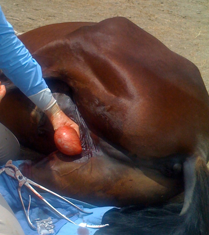
The castration procedure involves making a surgical incision at the scrotum and removing the testicles. The site is left open to drain and allowed to close naturally (it is not sutured closed).


Castrations performed by Dr. Devaney include:
  • Pre-operative tetanus vaccine (if necessary) [$15 value]
  • Pre-operative physical examination [$55 value]
  • General anesthesia, castration and pain control during surgery [$280 value]
  • Post-operative pain control for several days [$25 value]
  • Post-operative recheck examination [$35 value] 

Total value of $410 for $250 (plus call charge)

For the first postoperative day, the horse should be rested quietly in a stall and monitored closely for complications or problems. In the subsequent two weeks after the surgery, it is important to keep the surgery site draining, reasonably clean, and to moderate the swelling. This can be achieved by encouraging exercise twice daily or as needed. You may wash the surgery site with water (spraying gently with a water hose is acceptable) as needed to keep it clean. In addition, anti-inflammatory medication (e.g. Bute or Banamine) may be administered as directed by a veterinarian to help reduce discomfort and swelling.

For fly control, you may use regular fly spray on all intact skin. Catron IV spray works well around, but not in, the wound. For the wound itself, nitrofurazone spray (yellow) or alumimum spray (commonly called "silver spray" or brands like AluSpray or AluShield) can be applied on the wound.

After surgery, your horse may be incoordinated for several hours. Please do not ride or exercise the horse for 24 hours unless otherwise directed by your veterinarian. Please use caution in handling the horse as he may stumble or be incoordinated. If the horse drops his penis and fails to retract it for more than a couple hours, or it swells or becomes traumatized, seek veterinary care right away.

While the risks of serious complications are low, risks of castration include but are not limited to: swelling at the surgical site, excessive bleeding from the surgery site, infection at the surgical site, scarring or hydrocoele formation at the surgical site, damage to the limbs or body during anesthesia or postoperative recovery, evisceration through the inguinal rings, and anesthetic death.

If you feel your horse is exhibiting any signs which may suggest a complication is occurring, or if you have questions or concerns, seek veterinary advice immediately.

Post-mortem Exams

Has a beloved horse or other animal pet of yours passed away, and you wanted to know what caused the death?  A post-mortem examination is the way to find out.

I recommend a post-mortem exam on any unexplained or sudden death in any animal. Why? It can give real peace of mind if we find that the animal had a severe illness, such as cancer or heart disease. Also it helps us to know that the cause is not contagious, so that other animals living together would not be at risk. Finally, if the death was suspicious it can help us be reassured whether there was a toxin, foul play, or any other unusual factors involved. 

As with any test, a post-mortem is not perfect. Some things don't show up well on post-mortem exams and are better tested with antemortem blood tests, for example, such as cantharadin or oleander poisonings in horses. However many causes of death are apparent and having a post-mortem can help explain what happened to your beloved animal.

Here is a recent example from a fellow veterinarian, about a client whose dog died suddenly after surgery and taking pain medication. The vet and owner were worried the death might have been due to the surgery or a reaction to the medication.

 Last week I had a patient come in because he did not feel well early that weekend and the owner noticed a mass pop up at the sternum. In talking to the owner, Sam had some vomiting but felt better later that week. I examined him and everything was fine. I did note that the 3 cm mass at the sternum did not feel like your typical lipoma and every time I did a fine needle aspirate I would get blood. So I recommended the removal of the mass. I normally do surgeries on Tuesdays but because the man was going out of town, I decided to remove the mass the previous Friday.

Well Friday came around and removed the mass. The mass had like a large clot in the center of it and I sent it off for pathologic analysis. No issues and sent the animal home. Well Saturday night I get a call from the owner that Sam as not doing well and it all started after the Deramaxx tablet (pain medication). I did not see the call until the next morning. He called me the next morning to say Sam was barely breathing. I told him to take the dog to the e-clinic where he came in DOA (dead on arrival). Of course the man was very upset and wanted to know what happened.

I know we are super careful about recording parameters during anesthesia. We have charts that must be filled out by the techs every 5 minutes. I explained to him that I did not know what happened and recommended he have an autopsy done. I explained that I would pay for it and I would help us know if there was anything we missed. Couple hours later, I got a call from the University hospital stating that he was there for a necropsy (post-mortem).  I get a call from the pathologist stating that Sam bled out from a splenic tumor. He also had mets [metastatic tumors that spread] to the lungs. I got the biopsy report of that mass which came back hemangiosarcoma [malignant cancer of the blood vessels which often starts in the spleen and spreads from there].

I spoke to the man who thanked me for having him do the necropsy. He said it turned the whole situation from the worst possible outcome to knowing that there was nothing that anyone could have done. I also think in hindsight that the vomiting/lethargy episode early that week was probably an acute bleed.